The Hong Kong Minimally Invasive Brain & Spine Neurosurgery Centre has top specialists who treat complex neurological conditions caused by stroke, tumors, hernias, injuries, and traumas. Each day our skilled neurosurgeons perform dozens of complicated surgeries, saving people’s lives and helping them to restore their health. Thousands of patients go through the doors of our neurosurgery center every year which gives our excellent team of surgeons unparalleled expertise and ability to guarantee exceptional results to all of the patients.
If you are looking for an excellent neurosurgery clinic in Hong Kong, you have come to the right place. Fill in the online form to make an appointment with one of our specialist and start your journey to a healthy and happy life!
Initial stage of treatment is drug therapy for one or two months. The effectiveness of drugs in controlling the pain can help clinical diagnosis. But medications can only be a temporary solution, not a permanent cure for the true cause of trigeminal neuralgia that is: external factors compressing on the nerve and make it short-circuit.
Brain tumors can be subdivided into non-cancerous benign tumor and malignant cancerous tumor. If a tumor is originated within brain compartment ......
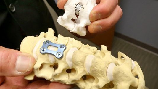
Tumors can develop in the vertebrae, nerves, and other tissue throughout your spine. Some spine tumors, such as astrocytomas, occur more commonly in children and adolescents......
Given the disease burden of strokes, prevention is an important public health concern. As stroke neurosurgeons, we do not want to treat stroke unless we are forced to do so for acute stroke ......
Brain tumors grow and compress normal brain tissue. Both benign and malignant tumors can cause swelling of the brain and raised intracranial pressure. Headache, dizziness....
中風有時也會被稱為腦血管意外(Cerebrovascular Accident CVA), 是影響頸部血管或大腦血管的一種疾病,從而干擾到大腦的供血或腦壓, 迅速領到大腦功能喪失。
為什麼腦中風的危險因素不能改變?
年齡 — 在55歲以後生活的每個十年有腦中風的機率大約增加一倍,。雖然腦中風是老年人中常見,很多65歲以下的人也中招。現在,由於生活方式的因素,年齡在30-50歲的成年人也受到影響。
遺傳(家族史) — 如果父母,祖父母,兄弟姐妹曾有腦中風的經驗, 你腦中風的風險是更大的。家族性趨勢水平通常與高血壓,高膽固醇血症和糖尿病有關。
性別 — 患腦中風是多見於男性多於女性。在多數年齡組,男性多於女性。然而,使用避孕藥和懷孕帶來的特殊風險,以女性腦中風患者,從而超過總數
中風前或心臟病發作 — 腦中風的人已經有很多次中風,一個人誰沒有的。如果你有一個心臟病發作,你腦中風的風險較太。
為什麼腦中風的危險因素是可以改變的治療或控制?
不良的飲食習慣 — 飲食高飽和脂肪,反式脂肪和膽固醇能提高血液中的膽固醇水平。飲食高鈉(鹽)可以促進血壓升高。常餐中添加多餘的熱量可導致肥胖。此外,包含五個或更多份水果,每天蔬菜的飲食可以降低腦中風的風險。
缺乏體力活動和肥胖 — 作為不活動,肥胖或兩者都可以增加你的高血壓,高血脂,糖尿病,心臟疾病和腦中風的風險。每天試圖得到一個總活動對大多數至少30分鐘。
定期休息時間和睡眠 — 在我們的臨床經驗,最近生病,休息不足,患者比較容易腦中風,睡眠模式觸發腦中風發作的風險也存在。
吸煙 (主動和被動) -近年來,有研究表明,吸煙是腦中風的重要危險因素。香煙煙霧中的尼古丁和一氧化碳在許多方面損害心血管系統。
藥物 — 使用口服避孕藥加上吸煙會大大增加腦中風的風險。濫用軟性毒品亦存在腦中風的風險。
濫用酒精 — 酗酒可導致多種並發症,包括腦中風。過量飲酒可引起腦中風。對於那些喝酒每天不超過兩杯的人士,男性的建議每天不超過一杯,非懷孕婦女最能反映科學酒精和腦中風風險的狀態。
高血壓 — 高血壓是腦中風的主要原因(佔腦中風風險的35-50%)和腦中風的最重要的可控危險因素。許多人認為,有效的治療高血壓是一個關鍵因為它可令腦中風死亡率的加速下滑。然而,由腦中風神經外科醫生一些特殊的臨床情況發現患者與預先存在的血管狹窄如頸內動脈狹窄,需要稍高的血壓水平進行調整,。
高血脂 — 高血脂引起動脈粥樣硬化,從而增加腦中風風險 。看來,低高密度脂蛋白("好"膽固醇)對腦中風的男人是一個危險因素,但需要更多的數據來驗證的婦女其效果。降脂藥物已經顯示了約15%,能減少腦中風的風險
糖尿病 — 進行家族病調查,糖尿病是腦中風的獨立危險因素。糖尿病增加腦中風的風險至3倍。許多糖尿病患者也有高血壓,高血脂和超重。這更增加了他們的風險。
頸動脈或顱內動脈 — 在你的脖子供應血液到大腦的頸動脈- 。頸動脈狹窄是引起動脈粥樣硬化的脂肪沉積形成斑塊縮小動脈管腔的疾病。因此,血流供應大腦受到損害,它可能會或可能不會引起症狀患者。有時小的脂肪沉積在動脈壁會移動,遠端流動和阻止腦血管,造成栓塞性腦中風。有時本病也取得了頸動脈是容易受傷(動脈夾層傷),或容易被阻塞血塊,從而導致嚴重腦中風。當動脈病發生在腦即顱內動脈狹窄的動脈內,這也將導致腦中風的類似方法。
外週動脈疾病 — 這是運載血肢體和器官的血管狹窄。它在動脈壁造成斑塊的脂肪建立。患有外週動脈疾病有頸動脈疾病或顱內血管性疾病,這引起了腦中風的風險較高。
心房顫動 — 作為常見於老年人,這種心臟節律紊亂引起的腦中風風險。心臟的上腔顫動,而不是有效地跳動,它可以供應血池和血塊。如果血塊脫落,進入在動脈導致腦,導致腦中風的血液和心房。那些與心房顫動每年有5%的腦中風風險,而這種風險是高於那些與瓣膜性房顫。[根據腦中風的風險,抗凝或抗血小板藥物是必要的腦中風預防
其他心臟疾病 — 人與某些類型的先天性心臟缺陷,心臟瓣膜病,冠狀動脈心臟疾病,擴張型心肌病(心臟擴大),心臟衰竭患有腦中風的人比那些心臟中正常的人風險較高。
中風是因為腦部動脈血管出現問題而起,三十歲後中風的發病率會以倍數增加。其實,中風的病變是有跡可尋的,透過中風風險評估,我們可清楚了解腦部血管的健康狀況,及早處理問題,減低中風風險。
香港大約有七成的中風個案屬於急性缺血性中風。症狀雖然沒有像出血性中風的來得突然`嚴重和迅急, 但缺血性中風的急性治療卻要比出血性中風治療需求更加快和緊急,因為如果可以在黃金八小時內迅速打通血栓塞的腦血管,缺血的腦細胞就可以重新獲得血液供養, 大腦細胞受損的程度可減至最輕, 病人獲得治愈及完全康復的機會就可大大提升, 有些病人甚至可以完全沒有中風的後遺症和傷殘。
傳統黃金3至4.5小時治療方案的不足
治療急性缺血性中風 (AIS)的傳統方案,是於中風症狀出現後的三至四個半小時內於靜脈注入溶血劑(組織胞漿素原活化劑/recombinant tissue plasminogen activator) (iv rtPA),亦即Activase。但病人往往因為送院太遲、或因診斷的延誤、 或因被歸類為不宜使用溶血劑,所以超過九成半的急性缺血性中風病人, 都不能從黃金3至4.5小時靜脈注入溶血劑的治療方案獲益。
此外,靜脈注入溶血劑rtPA的黃金3至4.5小時治療方案可能無法有效地 治療某類型的缺血性中風: 如中風是因為大血管閉塞, 或是因為過長血凝塊阻塞, 或是因為閉塞性動脈夾層, 或是因為血管創傷而引起的缺血性中風,靜脈注入溶血劑rtPA都可能無效。
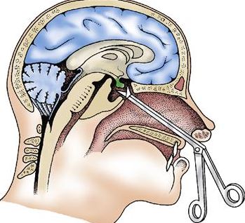
大腦通波仔,治療急性中風的黃金八小時
香港腦脊中風及痛症顧問中心 推出治療急性缺血性中風的嶄新「黃金八小時」概念: 腦內血管介入的微創手術(機械式吸取血栓術),正正填補了靜脈注入溶血劑rtPA的黃金3至4.5小時治療方案的不足。 病人可於中風症狀出現後的八小時內接受腦內血管介入的微創手術(機械式吸取血栓術),把血管再次灌通。就像心臟的通波仔微創手術, 專攻腦內血管治療的神經外科醫生會從腹股溝把微型導管放入腦血管,擊碎及吸走腦內血栓,把血管再次灌通, 令腦細胞重獲生機。腦內血管微創手術在大腦血管造影導引下,不但能於大腦栓塞的確實位置進行治療,高度準確地移除血栓。 有時候,如果神經外科醫生發港現病變和狹窄的腦動脈, 而其是造成中風的主要原因,醫生植入柔軟的金屬支架, 將大腦動脈重建。
鑑於中風的疾病負擔甚高,預防中風是一個重要的公共衛生議題。作為專門治療中風的腦神經外科醫生,我們並不想治療急性中風,因為治療急性中風的風險甚高, 但在緊急情況下, 為着拯救生命, 我們都被迫為之。但其實我們是希望提倡防止中風,促進中風風險篩查及中風預防性治療。
中風倖存者容易在未來再中風?
是 - 曾中風的人比正常健康的人有更高的再次中風個風險。如果你曾有心臟病發的病史,你再次中風個風險也是高較。
治療中風的真正成因,以防止短時間內再次中風:
採用傳统的醫療方法,大多數中風治療是盲目性的,無需檢索中風的真正原因而盲目下藥。現時專門治療中風的腦神經外科醫生則會以高品質的MRI造影或腦血管造影,以檢索中風的真正成因, 並把相應的中風風險移除,從而防止中風在短時間內再次發生。
中風患者能否成功地完全恢復健康,物理治療師是一個十分重要的角色。物理治療對中風的早期急救及之後的康復訓練十分重要, 即使嚴重中風令病人重度殘障或意識不清,物理治療亦應在中風後立即開始進行,以把握康復的黃金時機及減低中風的可致命併發症-----肺炎、深層靜脈血栓塞及褥瘡等等。
物理治療可以維持病人氣管及肺部的分泌物暢順排出,以減低罹患致命肺炎的風險。中風後,患者癱軟無力的肌肉在接下來的十數天會逐漸呈現繃緊的狀態,即是肌張力過高。如中風患者無法下床行動,小腿肌肉下就大有可能產生可致命的深層靜脈血栓塞(Deep Vein Thrombosis DVT) 的併發症。因此要預防DVT,就要替中風患者穿上具彈性的壓力袜子及定時進行大小腿肌肉按摩; 有時一些罹患DVT的高危病人, 更有可能需要服用抗凝血藥物,以減低患上致命DVT的風險。
在物理治療專注於矯正四肢和身體動作的同時,職業治療的大部份心思就放在協助患者重獲有用及實際的機能上,就以患者需要使用刀叉來吃肉為例,職業治療的主要目的,就是幫助中風患者盡量擴展他們的能力,使他們可以操作一般日常生活的工作,不但可以增强患者的獨立自主,也同時提升了他們的生活品質。
當中風患者情況隱定,職業治療師便會開始評估中風對患者所造成的損傷程度,並列出足以反映患者需要優先考量的狀況,以訂立其需要的治療計劃。
每個患者的期望,或其自主程度都不一樣,治療計劃也有很大的差異。例如,大部份上年紀的老人會希望恢復到可以獨立完成每天的日常瑣事便足夠,但年紀輕的患者也許就希望儘量重獲自己過去的許多技能。
大部份的中風患者都得參與特別的治療活動和工作,這些活動和工作能協助他們重新學習像如廁、梳洗、進食以及穿衣等必需的動作。這些活動和工作都有專門設計過的輔助器,以協肋進行上述的工作;例如,有一種特殊設計的淺平器具,它可以讓切割食物,以及把食物從盤子移到嘴裡等動作變得較容易。職業治療也會試着讓患者參與各種的嗜好和消遣活動,將情緒和心理上的壓力得以緩和,並讓患者覺得自己對家庭和社會仍有貢獻。
當疑似中風患者被送往公立醫院救治時,當值醫生會按病人情況而安排腦部的電腦斷層掃描(CT SCAN) 。CT是一個較經濟的初步影像檢查,但CT只能分辦出患者是屬於出血性中風 (佔30%的中風案例),不能詳细確診患者是否患上腦血管梗塞性中風(佔中風案例大多數的70%)。 CT 在腦血管梗塞性中風發生的首24小時內, 影像是可以完全正常的, 在醫生亦不能確診下, 病人就因此枉枉錯失了在黄金8小時內打通腦血管, 搶救大腦細胞及完全康復的機會。然而兩種中風: 出血性和腦血管梗塞性中風的治療迫切性及方針是截然不同的。
腦梗塞性中風就是大腦或頸血管受阻塞,血液無法流通,令腦細胞功能暫時受影響, 若時間拖延了, 大腦細胞就會死亡, 神經功能亦永久受損。
Accurate diagnosis of back pain isn't always easy. The human spine is very complex, so it can be difficult to pinpoint the exact cause of low back pain or other symptoms. Additionally, other factors, like depression, anxiety, or stress, can contribute to the symptoms of back pain. MRI scanning is safe and produces outstanding images of the spine, but, unfortunately, it doesn't show 'pain', therefore, in more complex cases of back pain it can often be very helpful to inject X-ray dye and local anaesthetic into the back to identify, and in some cases, treat, the painful structures.
History: Your doctor will ask you questions about your back problems, other related problems and your general health and past medical history. You may be asked to fill out a diagram that asks you where your symptoms of pain, numbness, tingling and weakness are occurring.
Physical Examination: Your doctor will examine your back for flexibility and range of motion. This often involves testing the strength of your muscles and your reflexes to make sure that they are still working normally.
X-rays: Your doctor will usually arrange a series of X-rays. X-rays show the bones, but do not directly demonstrate soft tissues, however if disc degeneration is present, the X-rays will often show a narrowing of the intervertebral disc spaces between the vertebral bodies, which indicates the discs have become very thin or have collapsed. Bone spurs, called osteophytes, can also form around the edges of the vertebral bodies and around the edges of the facet joints. As the disc collapses and bone spurs form, the space available for the nerve roots can also start to shrink.
X-rays are often performed sitting or standing with the individual bending forwards (flexing) and leaning back (extending) to demonstrate abnormal motion, known as instability.
 |
| Fig. 1. Intervertebral disc |
Magnetic Resonance Imaging (MRI): is a non-radiation technique for imaging the spine that uses a magnetic field. It can demonstrate soft tissue structures, such as the spinal cord, intervertebral discs (Fig. 1), etc as well as bone.
MRI is the single most useful test available for diagnosing spinal disorders.
MRI images are used to examine intervertebral disc height, hydration and a disc's ability to exchange nutrients with its vertebrae.
hong kong spine and pain centre has a special weight bearing MRI scanner which takes images with the individual standing up – this can be helpful, as spinal symptoms often present when one is standing. (Fig. 2)
 |
|
Fig. 2. The special weight bearing MRI scanner takes images with the individual’s positions: upright, tilting, & supine
|
Computed Tomography (CT) Scan: This uses a computer to produce detailed three-dimensional images of the body from a collation of cross-sectional X-rays. Of all the imaging techniques that are currently available, the CT scan is best able to produce images of bone.
Bone Scan: This procedure involves intravenously injecting a small quantity of a slightly radioactive marker into the patient's vein, and then running a scanner over the area of concern. The scanner detects the marker, which concentrates in any region exhibiting high bone turnover. A bone scan is used when there is suspicion of tumor, infection, or small fractures, i.e., conditions that all result in high bone turnover.
Myelogram: This test procedure involves injecting a radiographic contrast media (dye) into the dural sac surrounding the spinal cord and nerves, and then taking X-rays of the spine. This demonstrates the nerve roots very clearly. Although this is a very high definition investigation, it is rarely used since MRI offers similar information without the need for an injection.
After baseline imaging studies of X-ray and MRI, most problems can be diagnosed with confidence. However, certain conditions such as when presentation is atypical, multiple level disease, prediction of the post operative outcome, complex problems (e.g. failed previous surgery) and anatomical variants may need injection tests.
Provocative Discography (PD): Provocative Discography (also called Provocative Disc Injection Test) has been used as a diagnostic modality for back pain since the middle of the 20th century [1]. It is an intricate procedure in which many factors play into the production of consistent, reliable, beneficial data in assisting the doctor to identify both the source of a patient’s pain, and the best possible treatment. It is an operator-dependent procedure in which the spine surgeon must use good clinical judgement to ascertain whether or not the patient is a good candidate for discography, what useful information may be obtained, and which technique to use.
Discography is not only to show the disc, but to identify problematic, injured intervertebral discs by reproduction of the patient's usual pain complaint during the procedure.
An ongoing debate involves a comparison between magnetic resonance imaging of the spine, and the information obtained by discogram. MRI is an excellent imaging diagnostic tool that can differentiate between different parts of the disc. MRI can also be used to define details of the normal ageing process of the disc.
In single level disc disease, the pain origin is usually correlated to the MRI findings, however there is a 30% chance that the MRI findings are not typically correlated with the symptom.
In multi-level disc disease, the situation is even more confusing. Each individual abnormal disc may present differently.
To establish the relationship between the abnormal findings in the MR images and the clinical presentation, we need discography to help us to make an accurate diagnosis of the pain origin. Usually your doctor can also treat the pain by therapeutic injection at the same time in performing the discography.
The most common discography procedure is Transforaminal Epidural Steroid Injection.
The advantages of discography are:
You should not eat or drink for 2 hours before the procedure.
You will be admitted to day surgery ward of the hospital.
The procedure is done under local anaesthesia and you will be lying face down for 30 to 45 minutes (depending on how many levels and if additional procedures are required).
The level of injection will be confirmed by X-ray screening.
Your back will be covered with sterile drapes (Fig. 3).
Your doctor will use lignocaine (a local anaesthetic) to numb your skin.
Your doctor will pass a thin needle into the disc under X-ray guidance (Fig. 4).
Your doctor will inject contrast into the disc space to see if it reproduces your usual back pain (Fig. 5). This is called the Provocation Test.
Your doctor will be able to see the shape of the disc, and any tears and contrast leakage using X-ray screening.
 |
 |
 |
||
| Fig. 3. Setting in the operating theatre for discogram | Fig. 4. The needle inserted into the disc space through a “safe zone”, labeled as green. | Fig. 5. Intra-operative images of discogram |
The next step is the Relieving Test. Your doctor will inject lignocaine into the disc to see the pain relief response. We will record the change of the pain level one minute after the lignocaine injection. We might inject a normal disc for comparison if neccessary. However, we try to avoid this to reduce the further discomfort and potential disc degeneration of a normal disc after puncture.
Additional pain-relieving procedures, such as epidural steroid injection, is usually performed during the discogram.
Twenty to thirty minutes after the procedure, you can move your back to try to provoke your usual pain. You will report your remaining pain, (if any) and also record the relief you experience during the next week.
You may or may not obtain improvement in the first few hours after the injection, depending on if the disc injected is your main pain source. On occasion, you may feel numb, slightly weak or have an odd feeling in your leg for a few hours after the injection.
You may notice a slight increase in your pain lasting for several days as the numbing medication wears off before the cortisone is effective.
Ice will typically be more helpful than heat in the first 2-3 days after the injection.
You may begin to notice an improvement in your pain 2-5 days after the injection.
Occasionally, you may have some pain for longer than a week.
You may take your regular medications up to 2 weeks.
You may be referred for physical or manual therapy after the injection while the numbing medicine is effective and/or over the next several weeks while the cortisone is working.
Risks of Discography?
Risks of discography include discitis, neurologic and visceral injury, dye reactions, spinal headache and others. Spinal cord injury, vascular injury, pre-vertebral abscess, and subdural empyema have all been reported post-discography [4]. In 1995 it was recognized that the incidence of complications with discography were 0.15% per patient and 0.08% per disc [5].
The possibility that discography may cause persistent back pain since discography is generally performed in subjects with pre-existing back pain. Patients with significant emotional and psychological issues and chronic pain and those with disability claims account for more than 80% of those with persistent pain after experimental discography [6].
Selective Nerve Root Block (SNRB): This is injection test and/or therapeutic injection procedure for nerve root symptom (radiculopathy). This is done by injecting local anaesthetic and steroid on the nerve root under the X-ray guidance (Fig. 6). The equipment, pre-procedure preparation and technique are identical to discography.
 |
| Fig. 6. Intra-operative Fluoroscopy of Selective Nerve Root Block Right L5 nerve root. Left: Lateral view. Right: Antero-posterior view. |
Facet or Sacro-iliac Joint Injections: Local anaesthetic injections into these joints are used to evaluate these joints as sources of low back pain. Judging by the effectiveness of the anaesthetic in relieving pain, a specific joint(s) producing back pain can be identified.
Low Back Pain is one of the most common reasons for a visit to the doctor. MRI is an excellent investigation but it does not show which level is painful. Discography is the most accurate investigation for identifying the source of the pain. Due to the multitude of presentations of spinal pain, in many cases, without discography, a definitive diagnosis cannot be made.
Discography may prevent patients from undergoing unnecessary surgery.
In patients having an operation, post-operative success has been shown to improve with the use of preoperative discography, especially in cases of multi-level disc problems.
Address:
Kowloon Centre: Unit 1107, Grand Centre, 8 Humphreys Avenue, Tsim Sha Tsui, Kowloon (MTR T.S.T. Station A2 Exit)
Hong Kong Centre: 18/F, Euro Trade Centre, 13-14 Connaught Rd Central, Central, Hong Kong (MTR Central A Exit)
Contacts:
Tel:2367 6116
Fax:2682 5216
Hotline:
Tel:8107 1616
E-mail:
info@brainandspine.com.hk
Website:
https://www.brainandspine.com.hk
Find us on:
Consultation Hours (By appointment)
Monday - Friday:
10:00 a.m. to 13:00 p.m.
15:00 p.m. to 18:00 p.m.
Saturday:
10:00 a.m. to 14:00 p.m.
Sunday & Holidays: Closed


")